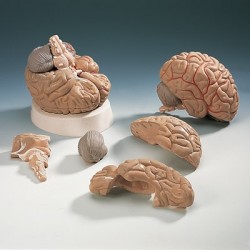
Trachea Section
Click Image for Gallery
This item is composed of 3 different models on the same base: the first one reproduces a 3X life-size cross-section of the human trachea; it can be longitudinally divided into two parts to show the inner anatomy. The second model is an enlargement of the anterior trachea wall cross-section; it shows all the different layers, from the tracheal cartilage to the epithelium. The third one shows the magnified pseudostratified cliated epithelium in great detail: it displays the typical ciliated cells and mucous cells.


